-
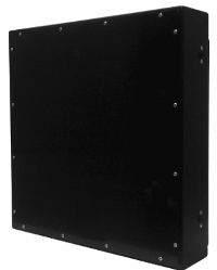
Digital x-ray systems based on multisensor technology (ССВ modules), used for direct conversion of x-ray photons (x-ray radiation) into video signal by means of several cameras with overlapping of fields and optical connection.
In the optical unit of the receiver x-ray beams are converted by intensifying screen into visible light, accumulated in video sensors. Each sensor processes relatively small field of view at intensifying screen, ensuring high resolution of the image. The more the number of video sensors in the optical unit the higher spatial resolution of diagnostic images is ensured by the receiver. Image quality can be improved with the help of zooming algorithms, selection of certain areas, setting of brightness and contracts, color inversion etc.
Key Features
Reliable modular design
High resolution of up to 4.6 lp/mm in the field of vision of 43 x 43 cm due to application of several sensors
Increased sensitivity by reason of high lens power (1:0.8) and high KE of sensors (>60%)
Much thinner and lighter as compared to one-camera systems.
Without cooling
Technological Advantages
High reliability and long operating time:
Electronic components, including sensors, are protected from direct x-ray action by means of lead and lead glass.
Quick image acquisition: less than 10 seconds between activation and deactivation of sensors after exposition. 1 year of receiver operation equals to 3 days of operation of sensors in video shooting mode.
Converting screen with the service life of more than 10 years and simple replacement.
Rigid body design.
Repairability due to modular design.
One of the best systems in terms of efficiency/cost parameters.
Simple and fast setting and calibration of the system.
High sensitivity due to careful selection of sensors, lens and screen.
Integrated function of post-processing of images to acquire diagnostic images of the highest quality.
Possibility of placement under any type of radioparent tables.
-
Technical specification
Receptor Type
Multiple photo-diode sensors array (PSA) with optical coupling
Sensors Number
120
Field of view, mm
244 x 304
Conversion Screen Type
DRZ-Std / CsI
Gad2O2S Green Fine
Radiography mode
Screen Pixel Pitch, µm
135x152
75 x 84
Spatial Resolution, lp/mm
3,7 x 3,3
6,7 x 6
Fluoroscopy / tomosynthesis / CBCT
mode 1
mode 2
mode 3
mode 4
Pixel Area
3600 x 3600
1800 x 3600
1200 x 1800
900 x 1200
Screen Pixel Pitch, µm
66 x 90
132 x 90
198 x 180
264 x 270
Nyquist frequency, lp/mm
7,5 x 5,6
3,7 x 5,6
2,5 x 2,8
1,9 x 1,8
Frame per second
2.7
5.5
20
30
Geometric Distortions
< 0,5%
Brightness Non-Uniformity
< 1% of full scale maximum after sensitivity correction inside active area
Scan Method
Progressive
A/D Conversion
16 bits
Non-responding Pixels Inside field of view
None
Data Output
Gigabit Ethernet
Wi-Fi IEEE 802.11n
Mechanical specification
Size, mm
365(v) x 345(h) x 70/751(d)
365(v) x 345(h) x 75(d) with extra sensors protectionWeight, kg
approx 4
Housing Material
Carbon fiber / plastic
X-Ray generator interface
X-ray exposure auto detection (AED)
Integrated
Primary Image Processing
Geometric Distortion Correction
Software implementation, based on test-objects patterns2
Non-Uniformity Correction
Full range, based on series of flat-field test objects images for the given output brightness levels
Receptor Power Supply
Power Input, V DC
24 VDC supply (1st stage)
+ battery-powered (2nd stage).
65W 230V (50 Hz) or 110V (60Hz) to 18VDC converter is provided separately and should comply all Council Directive 93/42/EEC requirements
Consumption, W
50 (max)
NC-01 digital panel
The goal of our company is
offer a wide range of goods and services at a constantly high quality of service
offer a wide range of goods and services at a constantly high quality of service


