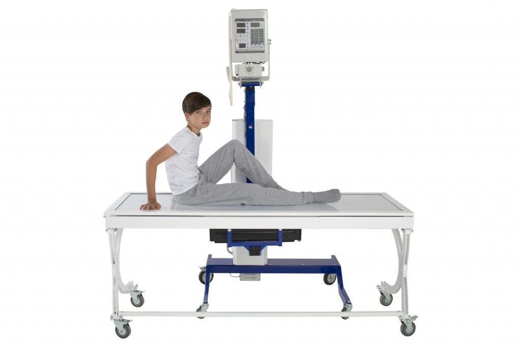
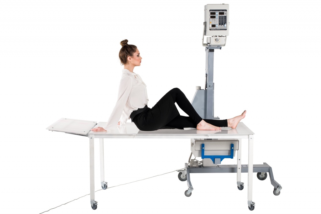
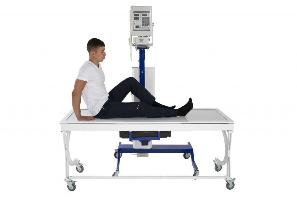
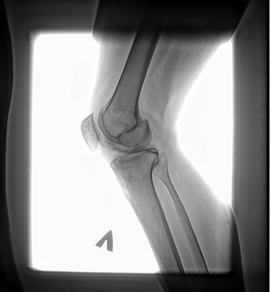
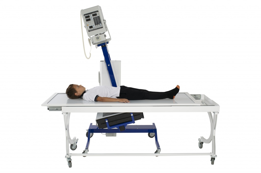
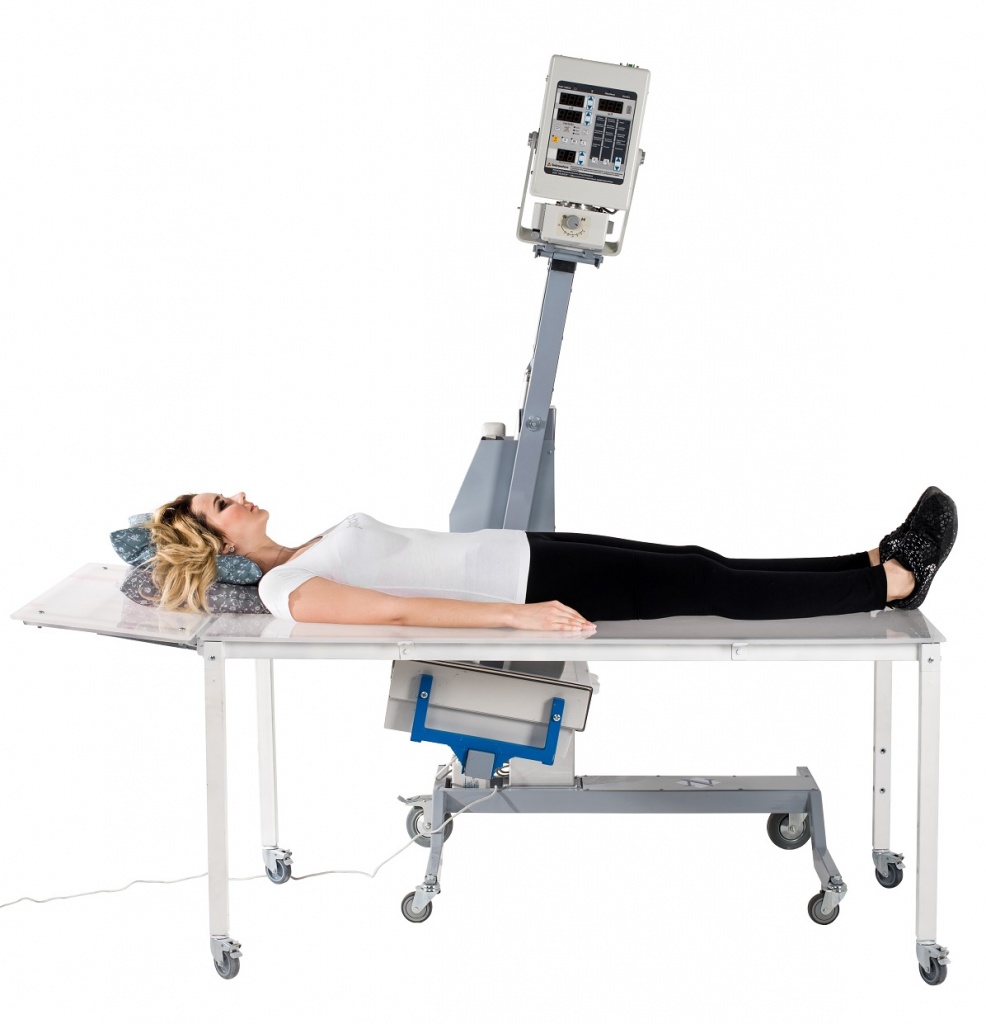
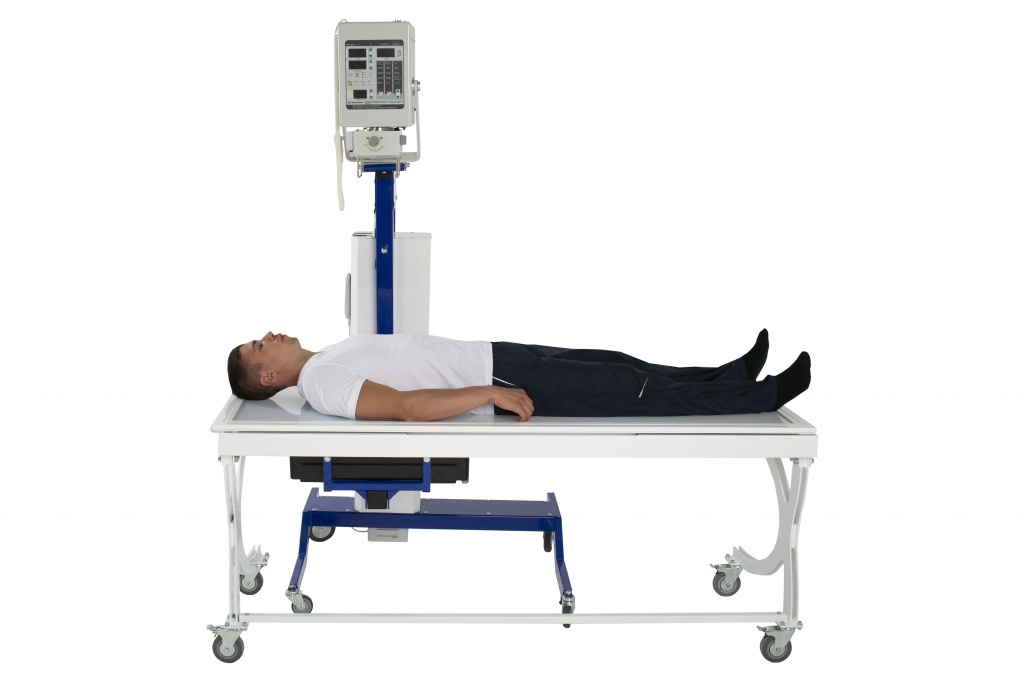
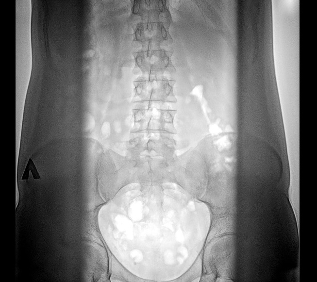
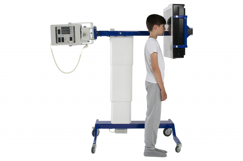
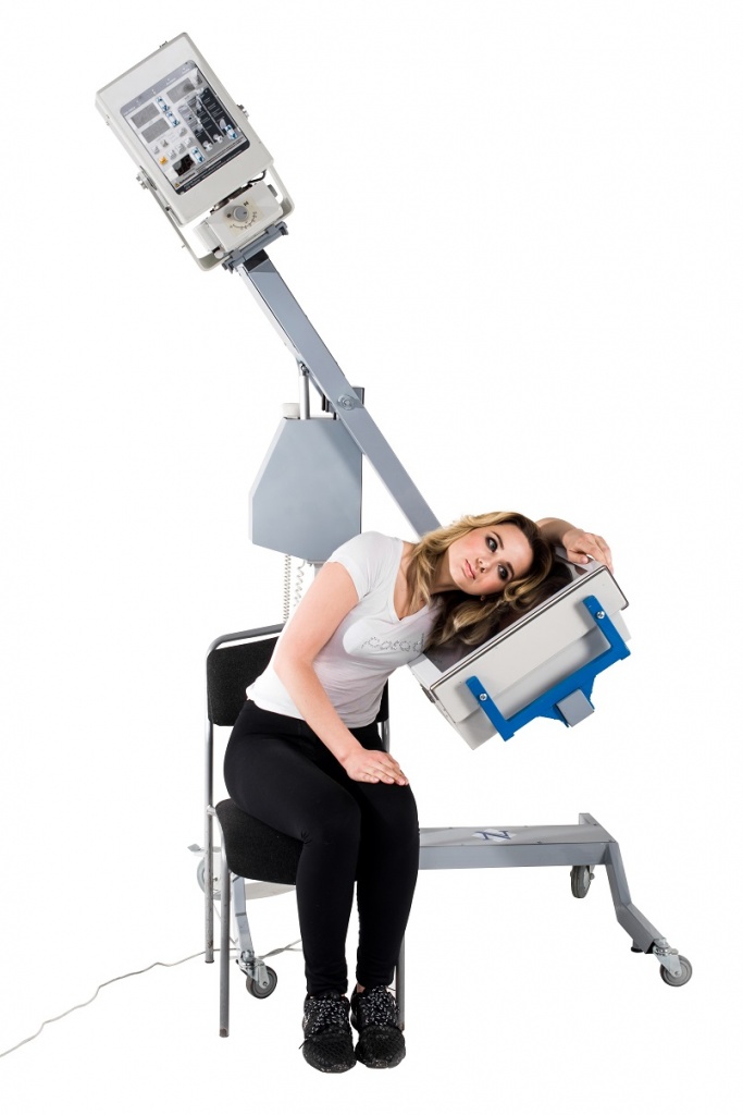
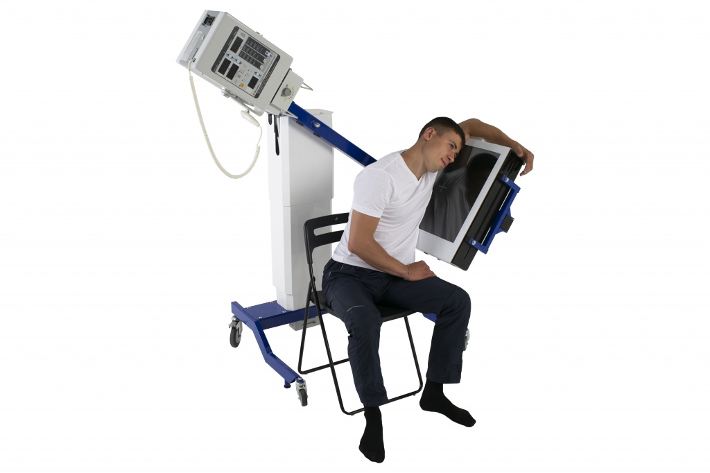
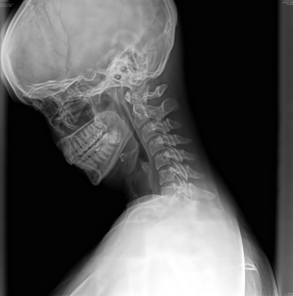
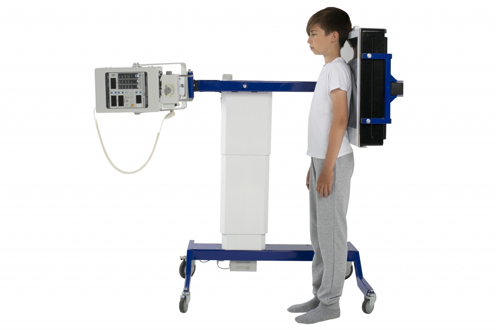
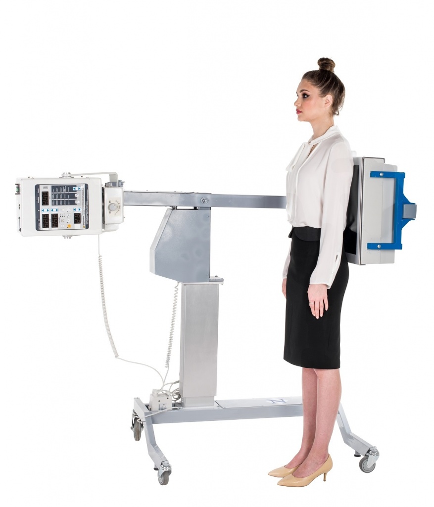
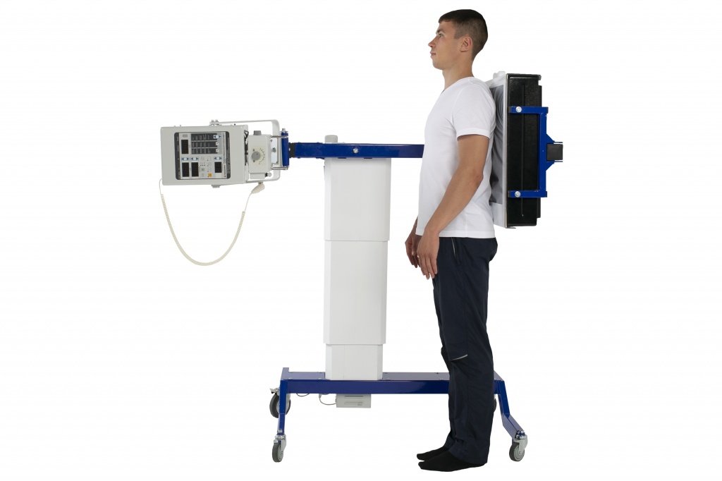
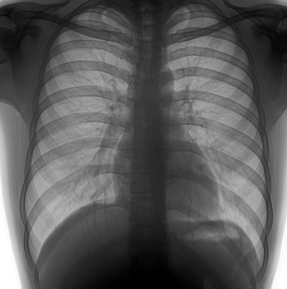
NOELSI company performs completing, assembling and adjustment of universal digital X-ray equipment set with lungs screening function (fluorography), which provides isometric image characteristics and complete convergence with the object of study.
Perfectly balanced swinging arm is on two-position rotary tripod on the wheels with safety blocking devices providing mounting of portable X-ray devices such as: DM-100P, DIG-360, EPX-F2800, PXP-60HF, EPX-F3200, PXP-100CA, meX + 100 and digital X-ray systems such as: PSA of NTs-01 model receiver, DR Rayence 1717 detectors, Carestream DRX-1 35x43 cm.
TECHNICAL SPECIFICATIONS OF UNIVERSAL X-RAY EQUIPMENT SET WITH LUNGS SCREENING FUNCTION (FLUOROGRAPHY)
| Power supply type | Inverter |
| Generator capacity, kW | 5.0 kW |
| Focal spots diameter, mm | 1.8x1.8 mm |
| Impulse frequency delivered to the rectifier of the power supply, kHz, minimum | 150 |
| Range of the anode voltage alteration of the tube, kV, minimum | 40-110 kV |
| Minimum voltage on the x-ray tube, kV, minimum | 40 |
| Critical voltage on the x-ray tube, kV, minimum | 125 |
| Supply. Phase. Frequency kHz, minimum | Single-phased /50-60 Hz |
| Step of the anode voltage alteration of the tube, kV, maximum | 1 |
| X-ray tube current range, mА, | 20-100 mA |
| Range mAs | 0.1 - 100 mAs |
| Voltage fluctuations %, maximum | 1% |
| AC power supply - voltage | 200-240 V ±10% |
| Maximum consumption of the current (А), maximum | 10А |
| Nominal grid resistance, Ohm | System automatically adjusts to the grid resistance via the voltage compensator |
| Kilovolt LCD indicator (kV) | available |
| Milliampere LCD indicator (mA) | available |
| Milliampere per second LCD indicator (mAs) | available |
| Density LCD indicator | available |
| “up/down“ buttons for kV values | available |
| “up/down“ buttons for mAs values | available |
| “up/down“ buttons for density values | available |
| Buttons for selecting automatic radiography parameters | available |
| Double detent button to turn on the emitter on a cable 5 m long | available |
| Illuminated collimator button | available |
| Buttons for selecting automatic radiography parameters site | available |
| Operational overall dimensions, mm | 568x268x233 |
| Built-in memory of imaging modes settings, total | 32 |
| Detector type | Multisensor photodiode conversion with optical communication and overlap |
| No. of sensors | 72 |
| Transducer type | Gd2O2S:Tb |
| Dimensions of the input working field, mm, minimum | (380 ± 10) х (380 ± 10) |
| Time of exposure, ms | from 10 to 200 |
| Resolution limit, line pairs per mm (lp/mm), maximum | 4.5 |
| Distortion and local geometric errors,%, maximum | 0.5 |
| Dynamic range, times, minimum | 250 |
| Exposure dose in the input plane, maximum: | 2.0 µGy (0.2 mP) |
| Enlargement coefficient of the image fragments on the screen | from 2 to 5 |
| Software functions | |
| storage of patients’ records by parameters: first name, last name, patient’s medical record No., year of birth, place of residence, date of examination, diagnosis in an electronic data bank; | available |
| image search in the electronic data bank for no more than 15 s; | available |
| description of X-ray diagnostic images using the “memory” of the electronic data bank and templates for possible diagnoses; | available |
| - synthesized image processing: | available |
| - adjustment of brightness and contrast; | |
| - scrolling, inversion and scaling; | |
| - changing the shape and size of the processing area of x-ray images; | |
| - designing the histogram of the distribution of image brightness elements; | |
| - contrasting the selected areas for image processing; | |
| viewing mode of up to 3 images simultaneously; | available |
| storing at least 500 stills on one optical disk | available |
| Overall dimensions of the tripod in transportable position, cm | 160x98x15 |
| Total weight, kg | 67 |
DESCRIPTION OF THE SOFTWARE
DigipaX is a German software designated to process medical imaging data.
Benefits
- Forwarding of the radiographs to a patient or a colleague on a data storage device or in electronic form (the original data is kept in the storage).
- Ability to connect to the archive. Once the images are uploaded, they are available in the network.
- Saving the image after post-processing. Virtual lead letters, summaries, automatic image conversions, background backup of the recent radiographs.
- Multi-user program unit allowing the images of several patients to be opened at the same time.
- Displaying several images in the full screen.
- Ability to generate tasks consisting of different studies.
- Visual selection of groups of organs for the tasks generation - virtual lead letters, summaries, automatic image conversions, background backup of the recent radiographs.
- Archive of documents (in the current DICOM format) with the ability to record documents along with DICOM images.
- Automatic loading of DICOM CDs.
- Composition of the various images (full leg).
- Ability to use several monitor displays.
- Magic Sharp Filters for contrast enhancement and sharpening (image sharpening function via the mouse click).
- Working with the PACS system.
Perfect devices compatibility (supports all modern DICOM printers).
Safety is a priority!
Protection is available.
Data is protected by a three-level security system.
Level 1: RAID system.
Automatic data mirroring to another hard drive. The system can still work once one hard drive aborts.
Level 2: Backup.
Automatic backup to external hard drive. Monitoring of drives in the digipax system: maintaining of the drive capacity; warning if the drive is missing.
Level 3: saving to DVD.
Manual saving to CD or DVD once a week.

